可注射的壳聚糖玻尿酸水凝胶用于软骨组织工程
在中老年群体中软骨损伤是导致残疾的主要原因。当前使用细胞和生物材料替换受损组织的组织工程方法正发展为新的治疗方式。蕞近美国加州大学洛杉矶分校生物工程学系的Min Lee教授团队在可见光下通过与核黄素(RF)光引发剂相结合的光交联法创造了一个含有甲基丙烯酸酯乙二醇壳聚糖(MeGC)和玻尿酸(HA)的可注射性水凝胶。
包含玻尿酸(HA)的可注射壳聚糖复合水凝胶适用于软骨缺陷的修复。这种复合水凝胶是在交联之前将HA溶解在光交联壳聚糖溶液中,包含有高分子量HA的交联壳聚糖形成了一个半贯穿的网络。研究者对于含有和不含有HA的各种引发浓度和辐照时间的壳聚糖水凝胶的凝胶形态和时间、其与细胞的相容性、机械强度和降解情况都做了相应的评估。此外还评估了软骨细胞的增殖和细胞外基质的沉积情况。
研究者首先用光聚合制备了壳多糖以及丙烯酸甲酯醇壳聚糖(MeGC)/HA水凝胶并对其进行表征。甲基丙烯酸酯基团在乙二醇壳聚糖上的取代度可通过MeGC的核磁共振氢谱计算而得到(Fig 1)。

Fig. 1.1 H NMR spectra of MeGC (400 MHz, D 2 O).
通过研究作为引发剂作用浓度MeGC水凝胶的凝胶时间(Fig 2a)可知,MeGC的凝胶时间迅速从90s降低到12s同时核黄素(RF)的浓度也从1.5增长至24 µM。在1.5 µM浓度的核黄素下加入HA能显著的将凝胶时间从90s降至45s。当RF的浓度增加到24µM时MeGC/HA复合水凝胶的凝胶时间进一步降低到11s。
研究者在冷冻状态研究了凝胶的内部形态(Fig 2b)。MeGC横截面的SEM图像显示了一个孔径大小在200~300µM的多孔微结构。而MeGC/HA复合水凝胶展示了更紧密的网络结构且其孔径大小在100~150µM,表明复合水凝胶具有更高的交联密度。用阿尔辛蓝染色观察未长细胞的复合水凝胶中HA的结合情况及其随时间的稳定性(Fig 2c),以没有HA的MeGC水凝胶作为对照组。结果表明交联之后的凝胶中存在HA。

Fig. 2. (a) Gelation time of MeGC and MeGC/HA (HA, 350 kDa) solutions as a function of RF initiator concentration. (Insets) Images of MeGC solutions with RF before (sol) and after (gel) irradiation. Gelation time significantly ( ⁄⁄ P < 0.01) decreased from 90 to 45 s with the addition of HA at 1.5 l M RF. There was no significant difference in gelation time between MeGC and MeGC/HA above 6 l M RF. (b) SEM images of MeGC and MeGC/HA (HA, 350 kDa) hydrogels. Hydrogels were prepared with6 l M RF and 300 s irradiation. Scale bar 300 l m. (c) Macroscopic images of MeGC and MeGC/HA (HA, 350 kDa) hydrogels stained with alcian blue after incubation in PBS at 37 ℃.
此外研究者对水凝胶的细胞毒性进行了测试。发现对于产生能够细胞封装和细胞培养的稳定凝胶需要蕞少40s的时间。在辐照时间是120~300s时细胞活性大约是87~90%,且在MeGC和MeGC/HA复合水凝胶中活性没有明显的不同。而当辐照时间延长到600s时会导致细胞活性的显著降低(Fig 3)。
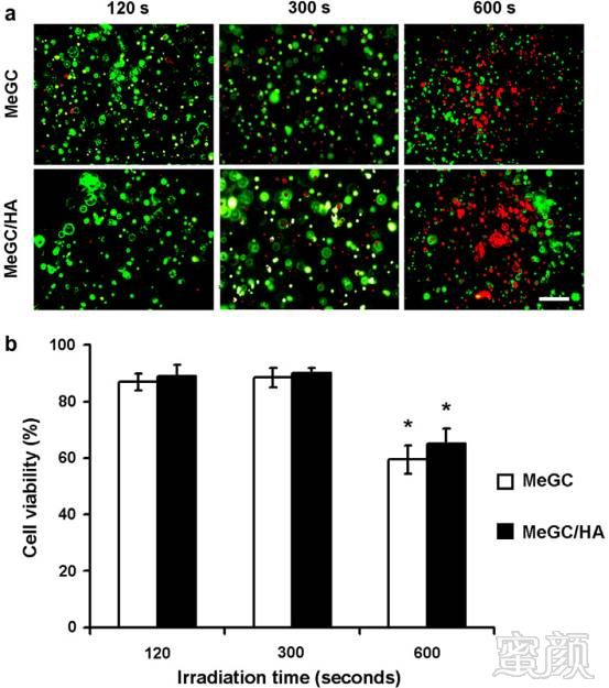
Fig. 3. (a) Live/dead fluorescent staining and (b) viability of encapsulated chondrocytes in MeGC and MeGC/HA (HA, 350 kDa) hydrogels polymerized for various irradiation times (120, 300, and 600 s). Original magnification 200?. Scale bar 100 l m. Two-way ANOVA F(2,12) = 3.89 and P = 0.14918 for interaction, F(2,12) = 3.89 and P = 0.00001 for irradiation time, F(2,12) = 3.89 and P = 0.61688 for HA addition. Tukey’s post hoc test confirmed that 600 s irradiation significantly reduced cell viabilities in MeGC and MeGC/HA ( ⁄ P < 0.05).
研究者还对在不同辐照时间和引发剂浓度下对存在和不存在HA的水凝胶的机械强度进行测定。当辐照时间从40s增加到600s时,MeGC的压缩模量显著增加,从大约为0.7增加到11KPa且当引发剂的浓度从1.5增加到15µM时会导致MeGC水凝胶压缩模量的显著上升(Fig 4a,b)。当RF的浓度等于或小于6µM时,含有HA的水凝胶会有更好的机械性能。而当引发剂的浓度增加到6µM时并不会导致MeGC/HA复合水凝胶机械性能的显著不同。
HA分子量对于水凝胶机械强度的影响通过使用低、中、高三种分子量的HA来表征,发现复合水凝胶的压缩模量随着HA分子量的增加而显著增加(Fig4c)且接下来只对RF的浓度在6µM以及照射时间在300s时复合水凝胶进行了进一步的研究。
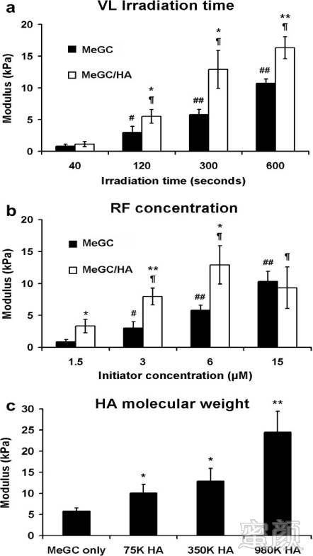
Fig. 4. (a) Mechanical properties of MeGC and MeGC/HA hydrogels polymerized for various irradiation times (40, 120, 300, and 600 s, 350 kDa HA, 6 l M RF): two-way ANOVA F(3,16) = 3.24, P = 0.00032 for interaction; F(3,16) = 3.24, P < 0.00001 for irradiation time; F(1,16) = 4.49, P = 0.00005 for HA addition. Tukey’s post hoc test,# P < 0.05 and ## P < 0.01, significantly higher than 40 s in the MeGC group; – P < 0.01,significantly higher than 40 s in the MeGC/HA group;/ P < 0.05 and ⁄⁄ P < 0.01,significantly higher than the corresponding MeGC group. (b) Mechanical propertiesof MeGC and MeGC/HA hydrogels polymerized at various RF initiator concentra-tions (1.5, 3, 6, and 15 l M, 350 kDa HA, 300 s irradiation): two-way ANOVAF(3,16) = 3.24, P = 0.00961 for interaction; F(3,16) = 3.24, P < 0.00001 for RF con-centration; F(1,16) = 4.49, P = 0.00035 for HA addition. Tukey’s post hoc test:# P < 0.05 and ## P < 0.01, significantly higher than 1.5 l M in the MeGC group;– P < 0.01, significantly higher than 1.5 l M in the MeGC/HA group; ⁄ P < 0.05 and⁄⁄ P < 0.01, significantly higher than the corresponding MeGC. (c) Molecular weight of HA (75, 350, and 980 kDa, 6 l M RF, 300 s irradiation): one-way ANOVAF(3,8) = 4.07, P = 0.00041 for HA molecular weight. Tukey’s post hoc test:⁄ P < 0.05⁄⁄ P < 0.01, significantly higher than MeGC alone.
研究者对MeGC水凝胶随时间的降解行为进行了测定(Fig 5)。由MeGC水凝胶的质量随着时间的损失可以观察到,在溶菌酶存在时,水凝胶的重量在42天的时候仍然是19%,而没有溶菌酶组在孵化时观察到有少量的质量损失。但复合水凝胶的重量在42天的时候仍然是50%且降解缓慢。
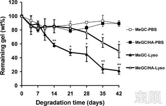
Fig. 5. In vitro degradation of MeGC and MeGC/HA (HA, 350 kDa) hydrogels after incubation in PBS in the presence or absence of lysozyme (10 mg ml ?1 ) at 37 ?C. Hydrogels were polymerized with 6 l M RF for an irradiation time of 300 s. Significant mass loss was observed in the presence of lysozyme (one-way ANOVA/ P < 0.05, ⁄⁄ P < 0.01).
为更进一步观察复合水凝胶在软骨组织工程中的可行性,研究者将软骨细胞封装在各实验组中且表征他们在长期培养过程中的细胞活性和增殖情况(Fig. 6a,b,c)。发现与纯MeGC水凝胶相比,含有HA的水凝胶对细胞增殖数目有显著的提高。

Fig. 6. (A) Live/dead fluorescent staining and (b) viability of encapsulated chondrocytes in MeGC and MeGC/HA (HA, 350 kDa) hydrogels after 1, 7, 14, and 21 days in culture,and (c) alamar blue assay showing proliferation of encapsulated chondrocytes cultured in hydrogels. Chondrocyte proliferation was significantly enhanced in the presence of HA (one-way ANOVA,/ P < 0.05, ⁄⁄ P < 0.01). Hydrogels were polymerized with 6 l M RF for an irradiation time of 300 s. Alginate (Al) hydrogels were used as the standard for comparison. Original magnification 100?. Scale bar 200 lm.
H&E染色显示封装的软骨细胞第壹天时在水凝胶中是作为个体细胞均匀分布的,在21天之后变成圆形且在MeGC/HA复合水凝胶中发现蕞高的细胞密度。由对比可知MeGC/HA复合水凝胶在缺陷处和细胞外基质周围的细胞集群中有更强的阳性蕃红染色和阿辛蓝染色。因此含有HA的MeGC复合水凝胶为软骨细胞提供了适宜的环境且保持了他们的形状,是一种潜在的修复软骨组织缺损的组织工程支架。
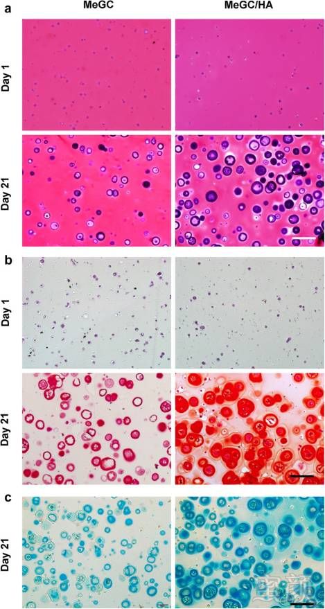
Fig. 7. Histological analysis of encapsulated chondrocytes in MeGC and MeGC/HA (HA, 350 kDa) hydrogels after 1 and 21 days culture. Polymerization was performed with 6 l M RF for an irradiation time of 300 s. Hydrogels were processed for (a) hematoxylin and eosin staining, (b) Safranin-O staining, (c) and alcian blue staining. Original magnification 200?. Scale bar 100 l m.
本研究由美国加州大学洛杉矶分校生物工程学系的Min Lee教授及其团队完成,于2012年8月27日在线发表于Acta Biomaterialia。
好消息!小编有个好消息告诉大家!近期,小编为大家在全国各正规医美机构争取了20个名额,大家可以低折扣抢购玻尿酸~目前还有8个名额可享有呦!别犹豫啦,碰碰运气~可以点击“在线咨询”,来进行名额预约~
